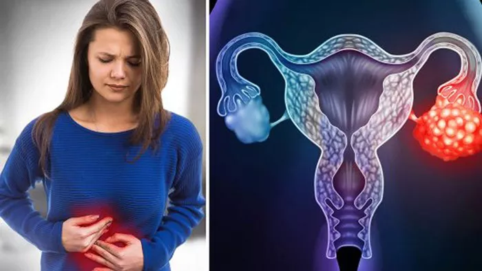
Uterine hematomas are collections of blood within the uterine wall or cavity. They can pose serious risks to both the mother and fetus, particularly during pregnancy. This article explores the causes of uterine hematomas, offering a detailed examination of the factors contributing to their development.
Unraveling the Ten Triggers of Uterine Hematomas
1. Trauma and Injury
Trauma to the abdomen is a common cause of uterine hematomas. This trauma can be due to accidents, falls, or physical abuse.
Blunt Force Trauma
Blunt force trauma, such as from car accidents or falls, can rupture blood vessels within the uterus. The impact and severity of the trauma often determine the size and extent of the hematoma.
Penetrating Injuries
Penetrating injuries, though less common, can also cause hematomas. Stab wounds or gunshot injuries can directly damage uterine blood vessels, leading to bleeding and hematoma formation.
2. Placental Abruption
Placental abruption is a serious condition where the placenta detaches from the uterine wall prematurely. This detachment can cause bleeding and hematoma formation between the placenta and the uterine wall.
Risk Factors
Hypertension, smoking, substance abuse, and trauma are major risk factors for placental abruption. A history of abruption in previous pregnancies also increases the risk.
Symptoms
Symptoms include vaginal bleeding, abdominal pain, and uterine tenderness. Diagnosis is typically confirmed through ultrasound and clinical examination.
3. Caesarean Section and Surgical Procedures
Surgical interventions, especially caesarean sections, can lead to the formation of uterine hematomas. During these procedures, blood vessels may be inadvertently damaged, resulting in postoperative bleeding.
Post-Surgical Hematomas
Hematomas can develop at the site of the uterine incision following a caesarean section. These can cause pain, fever, and potentially lead to infection if not properly managed.
Preventive Measures
Surgeons take measures to minimize bleeding during surgery, including meticulous techniques and ensuring proper hemostasis.
4. Invasive Prenatal Procedures
Invasive prenatal diagnostic procedures such as amniocentesis and chorionic villus sampling (CVS) can sometimes cause uterine hematomas.
Amniocentesis
Amniocentesis involves inserting a needle into the uterus to extract amniotic fluid. Although generally safe, there is a small risk of causing bleeding or hematoma formation.
Chorionic Villus Sampling
CVS involves taking a tissue sample from the placenta. This procedure carries a similar risk of bleeding and hematoma formation if not performed carefully.
5. Coagulation Disorders
Women with coagulation disorders are at higher risk of developing uterine hematomas. These disorders impair the blood’s ability to clot, leading to excessive bleeding.
Types of Coagulation Disorders
Hemophilia, von Willebrand disease, and thrombocytopenia are examples of conditions that can contribute to hematoma formation. Proper management during pregnancy is essential to prevent complications.
Management During Pregnancy
Close monitoring and appropriate treatments, such as clotting factor administration, are crucial to managing these disorders and reducing the risk of hematomas.
6. High Blood Pressure (Hypertension)
Hypertension during pregnancy, especially preeclampsia, can lead to the development of uterine hematomas. High blood pressure can damage blood vessels, making them more prone to rupture.
Preeclampsia and Eclampsia
Preeclampsia is characterized by high blood pressure and protein in the urine. If unmanaged, it can progress to eclampsia, which includes seizures. Both conditions can lead to placental abruption and hematoma formation.
Monitoring and Treatment
Regular blood pressure monitoring and antihypertensive medications are crucial for managing hypertension and preventing complications.
7. Abnormal Placental Attachment
Abnormalities in placental attachment, such as placenta previa and placenta accreta, can lead to uterine hematomas.
Placenta Previa
Placenta previa occurs when the placenta covers the cervix partially or completely, which can cause bleeding and hematoma formation, especially in the second and third trimesters.
Placenta Accreta
Placenta accreta involves the placenta embedding too deeply into the uterine wall, which can cause significant bleeding during detachment attempts, leading to hematoma formation.
8. Infections
Infections of the uterus, such as endometritis, can cause inflammation and damage to the uterine tissues, leading to bleeding and hematoma formation.
Endometritis
Endometritis is an infection of the uterine lining, often occurring after childbirth or surgical procedures. Symptoms include fever, pelvic pain, and abnormal vaginal discharge. If untreated, it can lead to hematomas.
Prevention and Treatment
Prompt antibiotic treatment is crucial to prevent complications, including hematoma formation. Proper post-surgical care and hygiene practices are essential to reduce infection risks.
9. Uterine Abnormalities
Congenital or acquired uterine abnormalities can predispose women to developing hematomas.
Congenital Abnormalities
Conditions like a bicornuate or septate uterus can disrupt normal blood flow and increase the risk of hematomas. These are typically diagnosed through imaging studies.
Acquired Abnormalities
Fibroids and adenomyosis are acquired conditions that can contribute to hematoma formation. Fibroids are benign tumors that cause abnormal bleeding, while adenomyosis involves endometrial tissue invading the uterine muscles, leading to inflammation and bleeding.
10. Assisted Reproductive Technologies (ART)
Assisted reproductive technologies, including in vitro fertilization (IVF), can increase the risk of uterine hematomas.
Ovarian Hyperstimulation Syndrome (OHSS)
OHSS is a complication of ART characterized by swollen, painful ovaries. Severe OHSS can lead to fluid leakage and hematomas in the uterus due to increased vascular permeability.
Embryo Transfer
During embryo transfer, catheter insertion into the uterus can cause minor trauma and bleeding, potentially leading to hematomas. Careful technique and ultrasound guidance can minimize this risk.
Case Studies and Real-life Examples
Examining case studies and real-life examples of uterine hematomas provides valuable insights into the practical aspects of diagnosis, management, and outcomes.
Case Study 1: Trauma-Induced Hematoma
A 28-year-old pregnant woman presented to the emergency department after a car accident. She was 24 weeks pregnant and experienced abdominal pain and vaginal bleeding. Ultrasound revealed a significant hematoma between the uterine wall and the placenta. Conservative management with bed rest and close monitoring was initiated. Over the following weeks, the hematoma gradually resolved without further complications, and she delivered a healthy baby at term.
Case Study 2: Placental Abruption
A 32-year-old pregnant woman with a history of hypertension presented with severe abdominal pain and vaginal bleeding at 30 weeks gestation. Ultrasound confirmed placental abruption with a large hematoma. Despite aggressive management, the patient required an emergency caesarean section due to fetal distress. The baby was delivered prematurely but recovered well in the neonatal intensive care unit. The mother’s condition stabilized after surgery, and she received close follow-up care to monitor her recovery.
Case Study 3: Coagulation Disorder
A 25-year-old woman with known von Willebrand disease became pregnant and was monitored closely by a high-risk obstetric team. At 20 weeks, she developed a small uterine hematoma detected during a routine ultrasound. Conservative management with bed rest and medications to support clotting was employed. The hematoma did not progress, and she continued with her pregnancy under close medical supervision, eventually delivering a healthy baby at 38 weeks.
Case Study 4: Invasive Prenatal Procedure
A 34-year-old woman undergoing IVF developed a uterine hematoma after a chorionic villus sampling procedure. She experienced mild abdominal pain and spotting. An ultrasound confirmed the presence of a hematoma. Conservative management with bed rest and close monitoring was initiated. The hematoma resolved without further complications, and the pregnancy continued to term with a successful outcome.
Case Study 5: Uterine Abnormality
A 30-year-old woman with a history of fibroids presented with vaginal bleeding and abdominal pain at 26 weeks gestation. Ultrasound revealed a submucosal fibroid and an associated hematoma. She was managed conservatively with bed rest and medication to control symptoms. Despite the challenges, she delivered a healthy baby via caesarean section at 37 weeks, and the fibroid was removed postpartum.
Conclusion
Uterine hematomas are a complex condition with various causes, ranging from trauma and surgical procedures to underlying medical conditions and anatomical abnormalities. Understanding the risk factors, diagnostic approaches, and management strategies is crucial for preventing and effectively treating hematomas. Regular prenatal care, careful monitoring, and appropriate interventions tailored to individual cases can significantly improve outcomes for both the mother and the fetus.
Healthcare providers play a vital role in educating and guiding patients, ensuring they receive the best possible care throughout their pregnancy journey. By addressing the underlying causes and implementing preventive measures, the risks associated with uterine hematomas can be minimized, promoting healthier pregnancies and safer deliveries.


